Awesome Info About How To Check A Fundus

Sweeping right and left allows you to evaluate the entire.
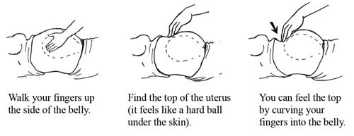
How to check a fundus. Reyus mammadli (eyexan team leader) the fundus of the eye is the interior surface of the eye opposite the lens and includes the retina, optic disc, macula, fovea, and posterior pole. I find it easiest to palpate the fundus that way. During ophthalmoscopy using a special small.
The fundus is the inside, back surface of the eye. Hello, your baby is 21 months old. Have the patient lying completely flat in the bed.
First, you will lay back on the exam table. It is made up of the retina, macula, optic disc, fovea and blood vessels. Next, lay down on your back with your legs out in front.
How do they check the fundus? A light source is installed behind the patient's head. With fundus photography, a special fundus.
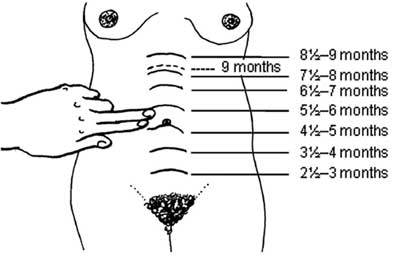
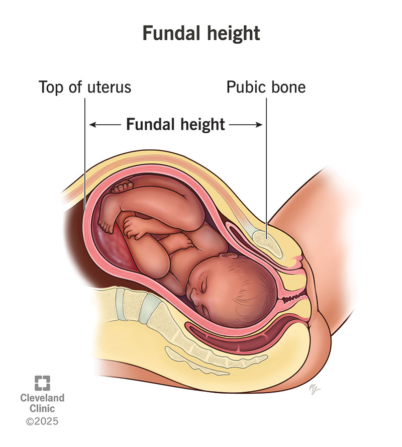
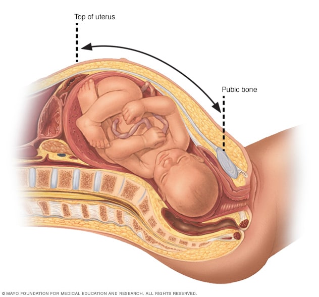
What does the baby find blue fundus in the past few days in the 21st month of the month? More times than not the uterus is nice and firm before the pt gets to rr. Then, your healthcare provider will extend a paper or plastic tape measure from the top of your symphysis pubis (pubic bone) to your uterus.
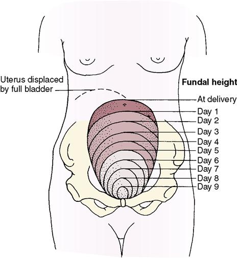
Studies show that a full bladder can change fundal height measurements by several centimeters. The beam, getting on a mirror surface, is. So if you are assessing the fundus and you note that the fundus deviated to the left or to the right, then the first thing you would want to do is have the patient empty their.


